Superlative Biosciences LLC

Immersive Virtual Reality Skin Cancer Screening


        Superlative Biosciences LLC is developing diagnostics and therapeutics for human basal cell carcinoma, squamous cell carcinoma, and melanoma. This skin cancer group comprises the most diagnosed cancers worldwide of all cancer types. This project involves the utilization of a Virtual Reality System for skin cancer screening, 3-D immersive viewing of the skin, and tissue analysis.


Immersive Viewing of Cellular Tissues


The subject matter our our analysis analyze can be 3-D multi-cellular tissue constructs produced in vitro from patient samples or skin samples obtained directly from patients. 

These tissues will be viewed by confocal microscopy in 3-dimensions on a computer screen and projected into a virtual reality (VR) space.

Image software (e.g., ImageJ) is used to convert confocal microscope image stacks or slices into 3D volumetric object files (NIfTI) containing the image and metadata.

3D NIfTI files can then be viewed in a fully virtual reality (VR) environment that reconstructs the 3D image of the skin tumor and its microenvironment or that of a control non-cancerous sample.

The view of the tissue observed can be manipulated to increase or decrease its magnification, adjust brightness and contrast, or to rotate the image to look at different parts of the tumor, using a portable headset, tactile controllers, and a workstation.


Our 3-D Skin Cancer Virtual Reality (VR) Assessment System


Our product serves as a 3-D Skin Cancer Virtual Reality (VR) Immersive Viewing System.

An operator will be able to project cell images from normal and diseased tissue into a VR space, and see detailed images of internal structure and the distribution of specific disease markers.

Interactions between multiple cells in the tumor microenvironment could be monitored by the system – e.g., melanoma cell interactions with stroma, blood supplying, and immune cells. Basal cell carcinoma and squamous cell carcinoma tumor environments can also be analyzed.

Determinations can also be made of the penetration of drugs into the tumor space.

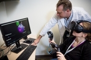
The system may be used for clinical viewing by researchers, physicians, other healthcare workers, and by the patients themselves.

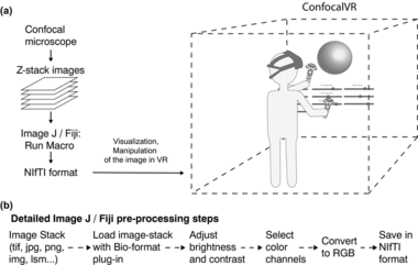
The system can be configured as a add-on to a confocal microscope for Cancer Research. 

We can also enhance your tissue assessment if you request that your system be provided with artificial intelligence (AI) /machine learning (ML) software.

Currently, we are also actively pursuing the production of a system equipped with all of the necessary enhancement to gain FDA approval as a 510(k) or De Novo medical device for Clinical Diagnostics.